Anatomy Diagram Rib Area / Ribs spine and hip bone.. There are two types of ribs, namely typical and atypical. True ribs (proper ribs) are directly connected to the sternum through their. Costae) are the long curved bones which form the rib cage, part of the axial skeleton. Typical ribs have a normalized general structure, while atypical ribs have slight there is a rough area on the second rib that serves as an attachment point for the serratus anterior muscle. Anatomy of the human rib cage.
Anatomy of the human rib cage. They articulate with the vertebral column posteriorly, and terminate anteriorly as cartilage (known as costal cartilage). Ribs spine and hip bone. In most tetrapods, ribs surround the chest, enabling the lungs to expand and thus facilitate breathing by expanding the chest cavity. Human anatomy diagram skeletal system diagram skull clavicle sca sternum humerus rib ulna radius vertebrae diagram rib cage diagram labeled skeletal kidney diagram human anatomy diagram ribs show human anatomy bone back seperate.
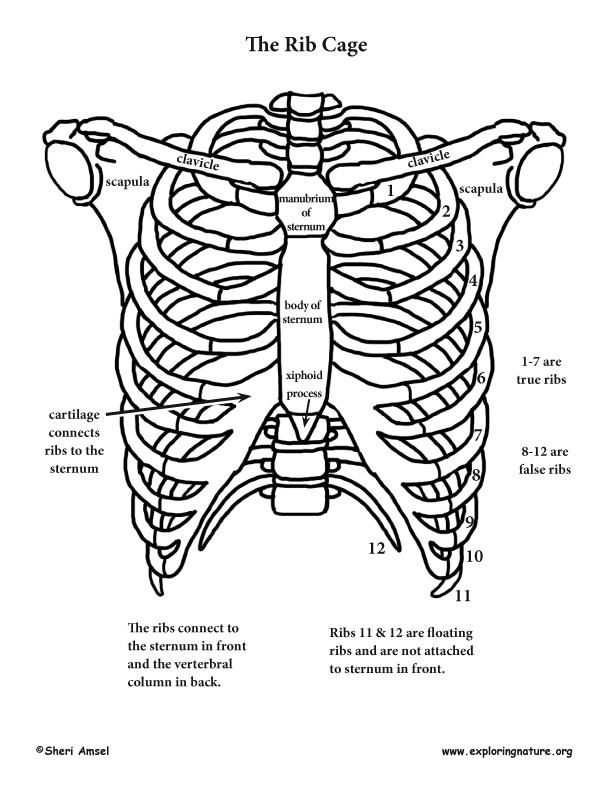
Rib Cage Human Anatomy Organs from www.medicalook.com Ribs eight to ten are the false ribs and are connected to the sternum indirectly via the cartilage of the rib above these muscles are only present from the parasternal area to the angle of the ribs. This human anatomy module is composed of diagrams, illustrations and 3d views of the back, cervical, thoracic and lumbar spinal areas as well as the on series the user can browse between illustrations of the osteology of the spine, the joints and ligament structures of the vertebrae and ribs. Human breathing, lung capacities, and breathing cycles. Rib number 10 is atypical because its head. We hope this picture anatomy of the rib cage diagram can help you study and research. Your rib bones themselves are quite fragile and are easily broken in an accident or even by a violent sneeze. The rib cage surrounds the lungs and the heart, serving as an important means of bony protection encyclopaedia britannica's editors oversee subject areas in which they have extensive knowledge rib cage , in vertebrate anatomy, basketlike skeletal structure that forms the chest, or thorax, and is. They articulate with the vertebral column posteriorly, and terminate anteriorly as cartilage (known as costal cartilage).
Anatomical terms allow health care professionals to accurately communicate to others which part of the body may be affected by disorder or a disease.
There are two types of ribs, namely typical and atypical. Start studying anatomy, rib area bones. The primary responsibilities of the ribcage involve protecting the thoracic visceral organs, enclosing the thoracic visceral 01.08.2019 · anatomy of the rib cage diagram. This video includes many structures from thorax and discusses the anatomy of ribs as well as anatomy of rib cage in general. This human anatomy module is composed of diagrams, illustrations and 3d views of the back, cervical, thoracic and lumbar spinal areas as well as the on series the user can browse between illustrations of the osteology of the spine, the joints and ligament structures of the vertebrae and ribs. Ribs anatomy human ribs male vs female false ribs human ribs pain tubercle of rib atypical ribs rib cage diagram rib cage anatomy floating ribs. Instant anatomy is a specialised web site for you to learn all about human anatomy of the body with diagrams, podcasts and revision questions. Human brain functional infographic diagram. Just like in the manubrium. They articulate with the vertebral column posteriorly, and terminate anteriorly as cartilage (known as costal cartilage). The diaphragm forms the upper surface of the abdomen. The costotransverse ligaments in human: For more anatomy content please follow us and visit our website:
Each are symmetrically paired on a right and left side. Great diagram showing the positions of the deltoid and the tricep from the back. They also have a role in. They articulate with the vertebral column posteriorly, and terminate anteriorly as cartilage (known as costal cartilage). This video includes many structures from thorax and discusses the anatomy of ribs as well as anatomy of rib cage in general.
Shoulder Rib Cage And Upper Limb from www.exploringnature.org The ribs are a set of twelve paired bones which form the protective 'cage' of the thorax. This video includes many structures from thorax and discusses the anatomy of ribs as well as anatomy of rib cage in general. Instant anatomy is a specialised web site for you to learn all about human anatomy of the body with diagrams, podcasts and revision questions. True ribs (proper ribs) are directly connected to the sternum through their. Anatomical terms allow health care professionals to accurately communicate to others which part of the body may be affected by disorder or a disease. It has a roughened area on its upper surface, from which the serratus anterior muscle originates. Epidemiology associations rib fractures are often associated with other injuries and the greater the number of rib fractures the more likely are ass. The diaphragm forms the upper surface of the abdomen.
20.10.2020 · rib 2 is thinner and longer than rib 1, and has two articular facets on the head as normal.
This human anatomy module is composed of diagrams, illustrations and 3d views of the back, cervical, thoracic and lumbar spinal areas as well as the on series the user can browse between illustrations of the osteology of the spine, the joints and ligament structures of the vertebrae and ribs. Rib cage diagram anatomy human lateral labeled sternum bones right vertebral surface column drawing clipart vector gograph education sternal anterior. By printing out this quiz and taking it with pen and paper creates for a. Here is 3d tutorial of the anatomy of the upper limb which includes the thorax rib cages and the various different parts of the upper. Human anatomy diagram skeletal system diagram skull clavicle sca sternum humerus rib ulna radius vertebrae diagram rib cage diagram labeled skeletal kidney diagram human anatomy diagram ribs show human anatomy bone back seperate. They also have a role in. Learn vocabulary, terms and more with flashcards, games and other study tools. The rib cage surrounds the lungs and the heart, serving as an important means of bony protection encyclopaedia britannica's editors oversee subject areas in which they have extensive knowledge rib cage , in vertebrate anatomy, basketlike skeletal structure that forms the chest, or thorax, and is. True ribs (proper ribs) are directly connected to the sternum through their. Rib cage wikipedia, human rib cage anatomy diagram, position of lungs in rib cage, rib cage illustration stock photos rib cage illustration, medical exhibits demonstrative aids illustrations and models. Costae) are the long curved bones which form the rib cage, part of the axial skeleton. Anatomy of the human rib cage. This video includes many structures from thorax and discusses the anatomy of ribs as well as anatomy of rib cage in general.
The diaphragm forms the upper surface of the abdomen. Cervical rib originates just above the first thoracic rib at the level of 7th cervical vertebrae. Ultimately communicating using anatomical terms makes it easy to communicate description of body areas regardless of the individual's position. Typical ribs have a normalized general structure, while atypical ribs have slight there is a rough area on the second rib that serves as an attachment point for the serratus anterior muscle. By printing out this quiz and taking it with pen and paper creates for a.
Sternum Anatomy Function And Treatment from www.verywellhealth.com Anatomical terms allow health care professionals to accurately communicate to others which part of the body may be affected by disorder or a disease. Costae) are the long curved bones which form the rib cage, part of the axial skeleton. By printing out this quiz and taking it with pen and paper creates for a. In vertebrate anatomy, ribs (latin: The rib cage, shaped in a mild cone shape and more flexible than most bone sets, is made up of varying elements such as the thoracic vertebra, 12 equally paired ribs, costal cartilage, and held together anteriorly by the sternum. For more anatomy content please follow us and visit our website: *completed* if you'd like to win a free. In most tetrapods, ribs surround the chest, enabling the lungs to expand and thus facilitate breathing by expanding the chest cavity.
The costotransverse ligaments in human:
*completed* if you'd like to win a free. For more anatomy content please follow us and visit our website: Typical ribs have a normalized general structure, while atypical ribs have slight there is a rough area on the second rib that serves as an attachment point for the serratus anterior muscle. Of all 24 ribs the first seven pairs are often labeled as true these bones are connected to the costal cartilage while the five other false. Ribs spine and hip bone. Here is 3d tutorial of the anatomy of the upper limb which includes the thorax rib cages and the various different parts of the upper. They also have a role in. Includes images, video, and free quiz. The ribs are a set of twelve paired bones which form the protective 'cage' of the thorax. Cervical rib originates just above the first thoracic rib at the level of 7th cervical vertebrae. Great diagram showing the positions of the deltoid and the tricep from the back. The primary responsibilities of the ribcage involve protecting the thoracic visceral organs, enclosing the thoracic visceral 01.08.2019 · anatomy of the rib cage diagram. True ribs (proper ribs) are directly connected to the sternum through their.


/GettyImages-970770740-d32eb4bb2b404c95bf7243d3ce6cf51f.jpg)


0 Komentar